From the National Institutes of Health
At a Glance
- A study in five volunteers undergoing surgery confirmed the existence of channels that may help drain waste from the brain.
- The results highlight the importance of ongoing research to boost the functioning of this waste-clearance system, called the glymphatic system.
Though less well-known than the body’s blood vessels, the lymphatic system is also vital to health. The network of lymphatic vessels threaded throughout the body removes dead cells and other waste from the bloodstream. It also helps transport the immune cells that fight infections.
It was once thought that the lymphatic system didn’t reach into the brain. But over the last dozen years, researchers have found a system of vessels containing cerebrospinal fluid in brain tissue in mice. These vessels appear to connect to the lymphatic system and to help clear toxins from the brain.
Studies suggest that age-related or physical damage to this brain waste-clearing system, called the glymphatic system, may contribute to the development of Alzheimer’s disease and other cognitive disorders.
Researchers have observed the real-time workings of the glymphatic system in mice. Studies using human brain samples taken after death found hints of such vessels. But to date, the existence of a functioning glymphatic system hadn’t been confirmed in living people.
In a new study, funded in part by NIH, researchers led by Dr. Juan Piantino from Oregon Health & Science University recruited five volunteers who needed surgery to remove a brain tumor. During this surgery, the volunteers received an injection of a dye called gadolinium into their cerebrospinal fluid. They then underwent MRI scans to track the passage of dye into the brain. Results from the study were published on October 7, 2024, in the Proceedings of the National Academy of Sciences.
One volunteer underwent MRI using a technology called T2/FLAIR at 12 and 24 hours after surgery, and the other 4 underwent T2/FLAIR imaging at 24 and 48 hours after surgery.
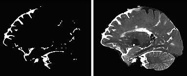
The scans showed cerebrospinal fluid flowing into the brain through distinct channels—along the perivascular spaces, the fluid-filled spaces that run alongside blood vessels in the brain. These findings match earlier imaging results seen in mice. Dye could also be seen moving from these spaces into the functional tissue of the brain.
“This shows that cerebrospinal fluid doesn’t just get into the brain randomly, as if you put a sponge in a bucket of water,” Piantino says. “It goes through these channels.”
Other studies have suggested that the glymphatic system may be most active during sleep. These new results support the importance of efforts to boost or repair the glymphatic system, such as improving sleep quality for people at risk for Alzheimer’s disease and other dementias.
—by Sharon Reynolds